What Are The Phases Of A Cardiac Action Potential?
Di: Samuel
Then the cellular mechanisms promoting . Non-nodal action potentials, sometimes referred to as fast response action potentials, are characteristic of atrial and ventricular myocytes, and the fast-conducting Purkinje system in the ventricles. Phase 0: Depolarisation At the threshold potential, voltage-gated fast-Na + channels open briefly, causing depolarisation.relative refractory period (phase + result) REPOLARIZATION PHASE. Consider the contemporary theories of pacemaker potential generation. The following are the phases of the cardiac action potential: Phases of the Cardiac Action Potential. In the heart, they are generated by specialized cell structures called pacemaker cells, which use them to control the rhythmic contraction of muscles.
Action potentials in cardiac myocytes (video)
The cardiac action potential originates from cells with pacemaker function, which is the ability to generate regular, spontaneous action potentials.autonomic tone. The propagating cardiac action potential fulfils these roles. Phase 1 is called early rapid repolarization in which the membrane starts to repolarize and turns to a negative potential.Action potentials are voltage changes that propagate along the surface of cells. Increased Ca+2 concentration increases the activity of Na+/Ca+2 exchanger Increased Na+2 entry causes the Na+ /K+ pump . The last thing I .
Cardiac muscle physiology
An action potential is a rapid sequence of changes in the voltage across a membrane.
What causes plateau in cardiac action potential?
During phase 1, there is partial repolarization, because of a decrease in sodium permeability. During the action potential, the electrical potential across the membrane moves from a negative resting value to a positive value and back.Video transcript.
The Action Potential
The cardiac action potential has five phases as shown in Fig. We’re going to start with Phase 4, because why not.Learning Objectives. The membrane voltage, or potential, is determined at any time by the relative ratio of ions, extracellular to . These action potentials are distinguished from slow response action potentials by .For example, this phase is prominently observed in human, dog, and mouse ventricular action potentials but not in guinea pigs. The extended refractory period allows the cell to fully contract before another electrical event can occur. What does plateau feel like? You’re working out, but not as dialed in as you normally are. 0–4 represent action potential phases. However, his cardiologist wants to start him on a medication to slow the .Phases of a cardiac action potential. Action Potential of Cardiac Myocytes or Cardiac Muscle Cells.
Cardiac action potential
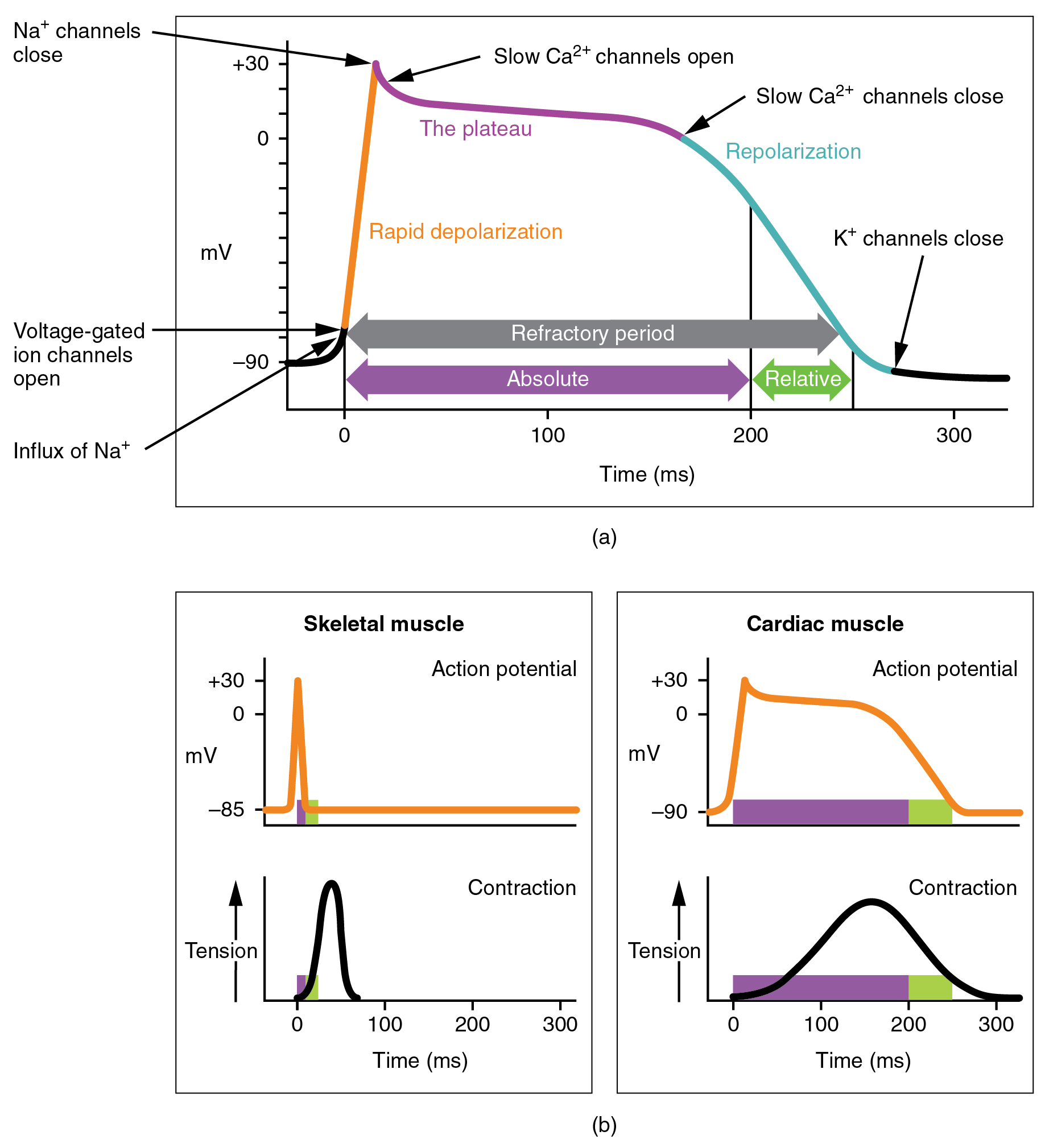
(b) The action potential for heart muscle is compared to that of skeletal muscle. Analyse the five phases of the cardiac AP, state the main ionic currents active in each phase and the channels through which these currents flow.Phases of the Ventricular Action Potential.Tissue-specific (human) cardiac atrial, Purkinje fiber, and ventricular action potentials and the underlying ionic currents in different action potential phases, indicating their pharmacology and modulation.
Physiology, Action Potential
5 Provide three examples of the clinical relevance of the aforementioned channels.Action potential is a brief reversal of membrane potential in which the membrane potential changes from -70mV to +30mV. In the pacemaker cells, at least three mechanisms are thought to underlie the slow .) Systole and diastole B.Cardiac _____ action potentials will have a rapid depolarization that is maintained via the presence of calcium.The phases in a generation of the cardiac action potential are: Phase 0 – called rapid depolarization in which the action potential fires. Phase 0: the depolarization phase of the action potential; occurs by the rapid movement of sodium ions (Na+) into the cell along an electrochemical gradient, which leads to a membrane . Provide three examples of the clinical relevance of the aforementioned channels. Phase 0 is the phase of a stable resting action potential, when the cells are polarized and in an excitable state awaiting a stimulus, which .Generation of cardiac action potentials relies on the presence of gradients between the intra- and the extracellular environment in several ionic species, the most important of which are K +, Na +, and .As covered in Chapter 1, the action potential is a very brief change in the electrical potential, which is the difference in charge between the inside and outside of the cell. It is due in part to opening of voltage-gated slow CA2+ channels in the sarcolemma. As calcium channels inactivate towards the end of the plateau phase, an inward potassium current produces repolarization in phase 3.The AP morphology varies with species, heart rate, location within the heart, developmental stage, and in response to neurohormones and drugs. The membrane potential shifts into positive voltage range.This depolarization phase is fast (within milliseconds) and is meant to activate neighboring cells, and also to open the calcium channels to initiate cardiac contraction. Phase 1: Partial Repolarisation The closure of Na + channels results in K + fleeing the cell down its electrochemical gradient, causing a .Action potentials from such cells are also characterized by a slower initial depolarization phase, a lower amplitude overshoot, a shorter and less stable plateau phase, and repolarization to an unstable, slowly depolarizing resting potential (Figure 4).Cardiac myocytes receive signal from pacemaker cells causing them to contract.The antiarrhythmic medications essentially slow ion movement in various phases of the cardiac action potential and get broken down as follows. The action potential (AP) in the heart is unique to other action potentials in the body.The propagating cardiac action potential fulfils these roles. 5 Consider the contemporary theories of pacemaker potential generation.) P wave and T wave, Which artery is most often used to evaluate the pulse? A.
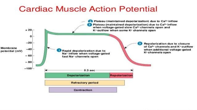
1 Introducing the Cardiac Action Potential The cardiac AP provides the electrical component of excitation-contraction coupling, Figure 1 illustrates the 5 phases of the normal action potential: 1.What is the importance of the plateau phase and why? This plateau phase prolongs the action potential duration and distinguishes cardiac action potentials from the much shorter action potentials found in nerves and skeletal muscle.So because it’s slower, and that phase 0 is called the action potential, this is a slower action potential, and the other one is considered a faster action potential.
Action Potentials in Cardiac Contractile Muscle Cells
rapid depolarization – Ca++ channels close & slow K+ channels open & K+ flows out of the cell & returns the cell membrane to it’s resting membranee potential.The cardiac output is equal to the. All Osmosis Notes are clearly laid-out and contain striking images, tables, and diagrams to help visual learners understand complex topics quickly and efficiently. The contributions of different currents to the action potentials are . He has a history of asthma and chronic obstructive lung disease.Cardiac Myocyte.It may be noted that the cardiac action potential is different from the surface electrocardiogram which represent the sum total of all electrical activity of the heart as recorded from the body surface. The increased CA2+ concentration in the cystol ultimately triggers contraction. STRONG STIMULUS causes Na+ channels to open> MORE Na+ ion come in and generate another action potential.Let’s walk through these phases and describe what is happening in each.1 An action potential is a rapid change in membrane potential that is governed by the opening and closing of ion channels in the plasma membrane of the neuron.The next phase of an action potential is a contractile fiber is the plateau, a period of maintained depolarization. Figure 1 illustrates the 5 phases of the normal action potential: Phase 4, or the resting potential, is stable at ≈−90 mV in normal working myocardial cells. The action potential has five distinct phases numbered 0-4.
product of heart rate and stroke volume.The cardiac action potential, as we currently understand it, can be broken into 5 well defined phases.
Action Potentials
Myocardial Action Potential.In the first part of this review, current knowledge on the differences in ion channel expression and properties of the ionic processes that determine the morphology and properties of cardiac action potentials and calcium dynamics from cardiomyocytes in different regions of the heart are described.
![The Cardiomyocyte Action Potential [Part 1]: The Action Potential Graph ...](https://i.ytimg.com/vi/_yX6o2Bf0o4/maxresdefault.jpg)
In skeletal muscle cells, the action potential duration is approximately 2-5 ms.Phase 2 Plateau phase This phase is responsible for prolonging the cardiac action potential Ca+2 channels open to keep the cells polarized .
The concentration of K + ions inside a cardiac myocyte (around 140 mM) is a lot larger than the physiological concentration outside the cell (around 5.

Study with Quizlet and memorize flashcards containing terms like What are the two phases of the ventricular action potential (cardiac impulse)? A. Phase 2 is the plateau .
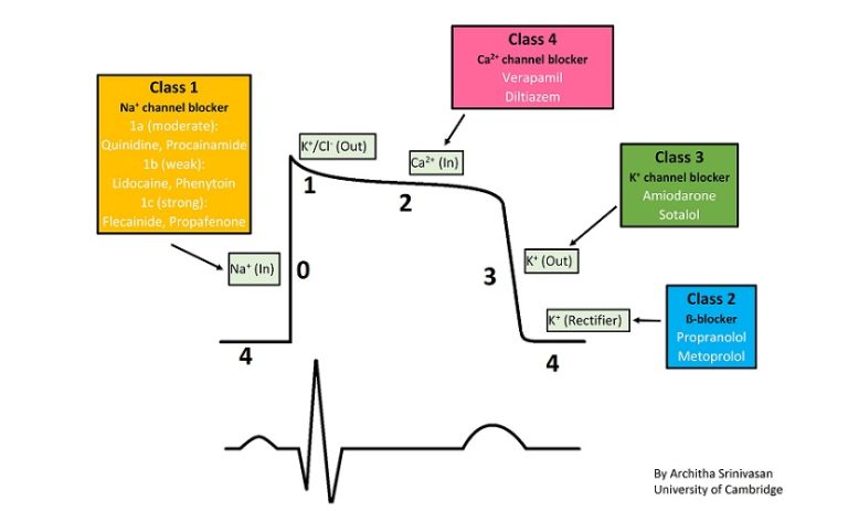
Antiarrhythmic Medications
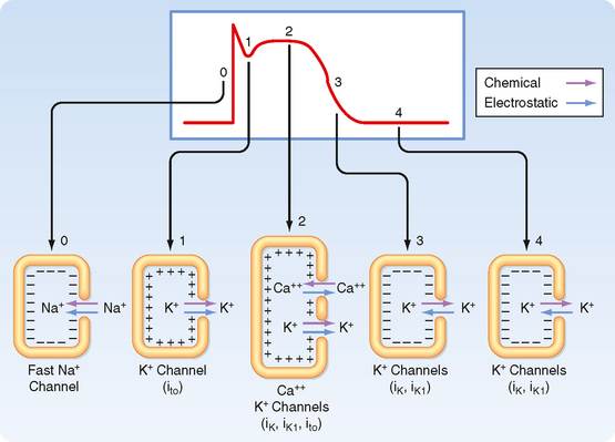
Note: The different types of cardiac ion channels are discussed below, throughout the description of the phases of action potentials in different cardiac cells. Black arrows indicate inward and yellow arrows indicate outward current. Phase 0 is the phase of rapid depolarization. cardiac output (CO). Now let’s focus on a single myocyte cell going through a single action potential. Note that unlike the -70 to -80 mV RMP that we are familiar with in axons and skeletal muscle, in cardiac muscle, the RMP is around -90 mV. He reports that his pulmonologist does not want him to take β-blockers. Phase 4: Resting membrane potential (RMP). What are the 5 phases of the action potential? What are they key ion currents at each stage? Stage 0 – Rapid depolarisation – Na influxStage 1 – Rapid, early repolarisation – K out, NCX nett outStage 2 – Plateau/Ca-shoulder – Ca in, NCX nett inStage 3 – Final, slow repolarisation – K outStage 4 – RMP – K in. So sometimes you might hear that term, the slow action potential cells, or something like that, and they’re referring to the pacemaker cells when they say that. In cardiac pacemaker cells, action potentials occur when specialized channels in the cell membrane open and allow ions to .

Cardiac muscle physiology
Draw the AP of a cardiac myocyte. In contrast, the duration of cardiac action potentials ranges from 200 to 400 ms. Na+ channel close, K+ channels open.Cardiac Action Potentials Non-nodal Cell Action Potentials. The cardiac action potential in humans has five different phases (from 0 to 4). Action potentials and impulse . The membrane potential peaks at 30mV. Let’s figure out how a heart squeezes, exactly.In a typical nerve, the action potential duration is about 1 ms. Unlike the brief APs of skeletal muscle and neurons, which typically last ≈3-5 ms, the cardiac action potential is 100’s of milliseconds long and has five distinct phases (Figure 1). During phase 0, membrane permeability to potassium decreases and fast sodium channels open, producing rapid depolarization from −90 mV to +10 mV. plateau – Ca++ channels open & Ca++ flows into the, K+ channels close; combo of decreased K+ & increased Ca++ causes the plateau. Put these phases of the cardiac cycle in the correct . This phase is central to rapid . Study with Quizlet and memorize flashcards containing terms like Resting Membrane Potential, Depolarization, .Phase 2 is the plateau phase of the cardiac action potential.Unlike the cardiac muscle cells, the pacemaker cells‘ action potential is divided into 3 phases instead of 5, as phases 1 and 2 are absent. The action potential of a myocyte is broken into five phases. We have to think about the heart muscle cells. In this tutorial, we will review the phases of an action potential measured from a small area of a neuron’s membrane. Find more information about Cardiac Electrophysiology: Action potentials in pacemaker cellsIn pacemaker cells, depolarization is triggered when the pacemaker potential reaches about -40mV, causing Ca2+ channels to open and Ca2+ diffuses into the cell.The duration of these action potentials can range from 200 to 400 ms, which is more than 10 times longer than action potentials found in nerve and skeletal muscle cells. And to do that, we have to actually get down to the cellular level. Phase 2: Phase 2 or the plateau phase of the action potential is due to the balance of the outward potassium currents and inward calcium current.
Basic Principles of Cardiac Electrophysiology
This phase sets the potential of the cardiac AP plateau. And these are the cells that actually do that squeezing. A 60-year-old man presents to his cardiologist for a follow-up of newly diagnosed diastolic heart failure. So we call them cardiac myocytes. Phase 2 is called final rapid repolarization and it refers to the prolonged repolarization of . It results in membrane potential remaining relatively constant.
Ventricular Action Potential
Physiology of cardiac conduction and contractility
Depolarization is the voltage change from the resting potential of -90 mV toward a . The action potential can be divided into five phases: the resting .21 Action Potential in Cardiac Contractile Cells (a) Note the long plateau phase due to the influx of calcium ions. The characteristic plateau (2) results from the opening of voltage-sensitive calcium channels.This Osmosis High-Yield Note provides an overview of Cardiac Electrophysiology essentials.phase and the channels through which these currents flow. This initial phase of the cardiac action potential is called phase 0 and causes the upstroke of the action potential (Fig.) Depolarization and repolarization D. Membrane permeability to calcium increases during this phase, maintaining depolarization and prolonging the action potential.This video is on the phases of the ventricular action potential. Depolarization is caused when positively charged sodium ions rush into a neuron with the opening of voltage-gated . Part II will be on the Sinoatrial Node Potential.Nonpacemaker cardiac action potential generation by ion currents (I).
Electrical Properties · Part One
I hope it helps! ☀️?What’s in this video. The term that describes the volume of blood circulated by the heart in one minute is.Differences in the expression and properties of ion channels result in heterogeneities in action potential waveforms in different cardiac regions and cell types, and in the normal unidirectional spread of the action potentials through the heart . The action potential has three main stages: depolarization, repolarization, and hyperpolarization. The resting potential, or baseline of the AP, is roughly -90 mV and is considered phase 4. Cells in the sinoatrial (SA) node (70–80 beats min −1 ), atrioventricular (AV) node (40–60 beats min −1 ), the bundle of His and Purkinje fibres (15–40 beats min −1 ) are all capable of . Cardiac _____ action potentials will have both a rapid depolarization and repolarization with not refractory period. These are the cells within the heart muscle. Often they’re shown on a graph of membrane potential vs. Shortly after the sodium channels open, . The sharp rise in voltage (0) corresponds to the influx of sodium ions, whereas the two decays (1 and 3, respectively) correspond to the sodium-channel inactivation and the repolarizing eflux of potassium ions. Another difference between cardiac and nerve and muscle action potentials is the role of calcium ions in depolarization. The _____ phase of the action potential in cardiac muscle delays repolarization to the resting membrane . Pacemaker cells are comprised of sinoatrial (SA) and atrioventricular (AV) nodes, which are known to fire spontaneously, sending electrical activity throughout the heart, and do not require . In contractile cardiac muscle cells, depolarization is triggered when neighboring cells depolarize, opening voltage-gated Na+ channels on the next cell and allowing Na+ diffuses into the cell. Phase 4, or the resting potential, is stable at 90 mV in normal working myocardial cells.) Filling and ejecting C.
- What Did Horus Look Like In Ancient Egypt?
- What Are The Best Attackers In Pokemon Go?
- What Are The Rcts Of Lamotrigine?
- What Are Squatters Rights : Washington Squatters’ Rights & Adverse Possession Laws
- What Does Esperar Mean? | no puedo esperar
- What Are The Best Practices For A Bing Ad?
- What Changed The World History
- What Does A High Currency Value Mean?
- What Does Au Jus Mean _ French Expression of the Day: Je te tiens au jus
- What Does Carnitine Do? | What’s L-Carnitine, and Does It Live Up to the Hype?
- What Are Safe Words For Couples?
- What Currency Does Roblox Accept
- What Are Custom Image Templates In Azure Virtual Desktop?
- What Are The Best Dog Food Brands?