Dog Leg Anatomy Diagram – Dog Leg Anatomy: Rear Leg and Front Leg Dog Anatomy
Di: Samuel
Other osteological features from dog ulna.Step 1: Preparation is Key. For the field practice, you might have a good piece of knowledge on the back, forelimb, hindlimb, and abdomen muscle anatomy from a cat. It sends an auricular branch to the lateral collateral . You will find great similarity in the osteological features of the cats’ hind leg bones with the dogs. The ilium is large and prominent in canines. Again, the muscles are also essential as most vessels and nerves pass along or within them. As the pace of veterinary advancement accelerates, even the most experienced veterinary teams are challenged to keep up with all the changes that impact their practice.
Dog Anatomy Guide
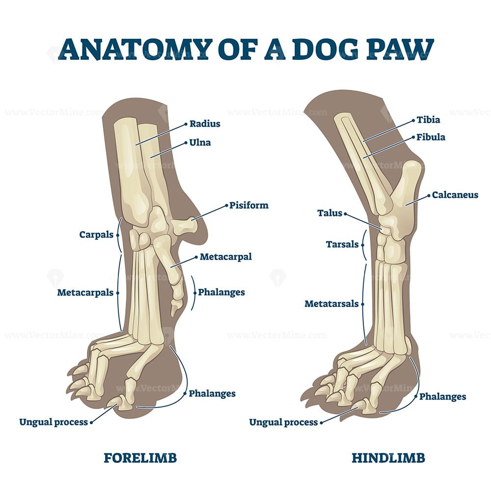
Here, in the dog (canine) spleen labeled diagram, I tried to show and identify the different surfaces and borders. Digital pads (4 pads that are located directly under the toes) Metacarpal pad (A large pad that is located directly under the digital pads) Dewclaw (A vestigial digit) Carpal pad (A tiny pad near the wrist)If the dog takes short, mincing front steps, or has to lower her head in order to extend her front leg to prevent it from colliding with the rear one, you know to take a closer look at her front .Leg Bones Anatomy, Diagram, and Function – Healthline. This nerve runs directly distal, obliquely crossing the lateral head of the gastrocnemius muscle.Dog legs (both front and back) are prone to injury in the short and long term since they spend so much time on them. Bony prominences are readily identifiable: these include the cranial dorsal iliac spine, the greater trochanter and the ischiatic tuberosity. Again, most of the .
Dog Hip Anatomy
Canine Bone Specifics. Some fascias, tendons, ligaments, joints were labeled. Internal dog anatomy organs.These include the head, ears, eyes, nose, mouth, neck, tail, legs, and paws.The cow leg anatomy consists of bones, muscles, nerves, and vessels.Dog leg anatomy with the labeled diagram – bones, joints, muscles, and vessels : Cow leg anatomy – bones, muscles, joints, and vessels from fore and hind limbs: Cat leg anatomy with the diagram – bones, joints, and muscles: Syndesmology or Arthrology of the Animals. The Atlas contains illustrations of the most . Dogs have knees, much like people, where the tibia and fibula meet the femur (where the lower leg meets with the thigh in dog hind leg anatomy). The femur of a horse is massive, and you will find an extra third . They do not stretch. Bones are the hardest and main component of the cow leg structure. Name * Email * Comment.
Complete guide to Dog joint anatomy
Dog leg muscle anatomy with the labeled diagram; But, you may also learn a little about these elbow joint muscles from this article (let’s see the muscles of the elbow joint section). Toe Bones (Phalanges of the Foot) What do the Digit Bones do, and Where are They Positioned . The head of a dog is one of its most distinguishing features.2021 Ultimate Guide to Dog Anatomy. The leg of a dog’s pelvic limb consists of the tibia and fibula .I hope the below-mentioned articles might help you to know these (bones and muscles) from the dog’s pelvic limb – Dog leg anatomy – bones, muscles, and vessels with the labeled diagrams, Again, the following article will also help you to get a basic idea of the different joints from the dog’s pelvic limb – Dog joints anatomy with the labeled . Add a comment Cancel reply.Cat hind leg anatomy.
Dog Liver Anatomy
In anatomy, this joint is called humeral-radial-ulnar. We can divide the canine skeleton into three main sections: Axial skeleton: skull, spine, ribs and sternum bones. dewclaw – the tiny, useless, fifth claw – located on the inner part of the leg above the other toes. But you will also find specific joints from the animal’s forelimb . Gluteal lines are not prominent in horse hip bone #2. For more information, consult the Bibliography, refer to prescribing information on specific drugs, or call Hill’s Veterinary Consultation Service at 1-800-548-VETS (8387) or e-mail vet_consult@HillsPet. A pregnant female dog’s . Tendons are tough bands of connective tissue made up mostly of a protein called collagen. Here is a list of anatomical structures that make up a dog’s paw: Claws.Dog pelvic bone anatomy. Popular Articles.
Dog Anatomy
A female dog’s reproductive system has similar organs as a human’s. carpals – the wrist, the bones of the pastern joint. The muzzle (foreface) comprised of the upper and lower jaws. Reproductive anatomy plays a role in sexual pleasure, getting pregnant, and breastfeeding. Dogs are domesticated descendants of the wolf and belong to the canine species. The urinary system helps rid the body of toxins through urination (peeing).
Components of the Musculoskeletal System in Dogs
Tendons attach each end of a muscle to a bone. ECVDI, Utrecht, Netherland) were categorized topographically into seven chapters (head, vertebral column, thoracic limb, pelvic limb, larynx/pharynx, thorax . Now, I will provide again the canine spleen anatomy labeled diagram that might help you to memorize the summary of it. Before embarking on our quest to locate the vulva, it’s vital to ensure maximum comfort for both you and your furry friend.
Anatomy of a Dog’s Paw with a Labeled Diagram
You will find 7 alveolar sockets on the posterior body of the mandible for cheek teeth (molar and premolar).
Label Dog Anatomy Printout
Additionally, dogs have a small bone inside the front leg called the accessory carpal bone, which humans don’t possess.Dogs exhibit extreme variations in body weight, shape and size.
Canine myology: normal anatomy
Table 1 and the diagrams provided in this article might help you locate the superficial segment of the cephalic vein from the dog’s thoracic limb. Again, a dog’s thigh is represented by the femur and associated sesamoid bones of the stifle joint. I hope you got the basic idea of these structures from the hind leg earlier. Here, I will focus on the external features and some of the internal features of the cats’ paws. This short post will try to cover thedog leg anatomyin detail. In dogs and other quadrupeds, as in humans, the movements of this joint are flexion and extension. In the dog the tuber coxae has two prominences; the cranial and caudal ventral iliac spines and although not usually visible, both are readily palpable.Anatomically, the term leg means the part of the hind limb that extends from the stiffle joint to the hock joint (knee to ankle or tibia and fibula bones region). Make sure your dog is in a comfortable position, either lying down or standing still. Here are presented scientific illustrations of the canine muscles and skeleton from different anatomical standard views (lateral, medial, cranial, caudal, dorsal, ventral / palmar. The hindlimb skeleton of the canine includes the pelvic girdle, consisting of the fused ilium, ischium, and pubis, and the bones of the hindlimb.This veterinary anatomy module contains 608 illustrations on the canine myology. brisket – the chest of the dog.This module of vet-Anatomy is a basic atlas of normal imaging anatomy of the dog on radiographs. Background: Anime is a type of Japanese animation that typically consists of high-quality, story-driven TV shows and movies.

If you’re an aspiring veterinarian, odds are you are going to see more than your fair share of injured knees over the coming years. – **Muscles:** The muscles in the dog’s leg, such as the quadriceps, hamstrings, and calf muscles, play a crucial role in movement and stability.
Dog Leg Anatomy: Rear Leg and Front Leg Dog Anatomy
Horse anatomy leg bones.Canine Anatomy The Dog. The main parts of the female anatomy can be broken up into outside . Femur (2): The femur or thigh bone is the longest and strongest bone in the body.Female anatomy includes the internal and external structures of the reproductive and urinary systems. We’ll break down the anatomy and function of the upper leg, knee, lower leg . The ears can be upright or floppy, and they come in a variety of shapes and sizes. The hindlimb has gluteal, perineal, thigh, knee or stifle, crural, tarsal, metatarsal and phalangeal regions. The labeled diagram shows the right lobe, left lobe, . The female dog anatomy external organ is the vulva, which opens to the vagina. I think you learn the basic idea on the cat leg muscle anatomy from this article. The bones implicated have contacting areas that resemble a hinge. In the pelvic dog girdle, you will find the ilium, ischium, pubis bones. The eyes are usually round and can be brown, blue, or green.
Dog Anatomy from Head to Tail
Also called the carpal joint, made of several (two rows) short bones. Here, I will show the course of the cephalic vein from the digits to the cranial . labeled joints anatomical limb. There is a trochlear notch (semilunar notch) in the cranial aspect of the olecranon process of ulna bone that helps .
Appendicular = forelimb bones + hindlimb bones. Luxating patella, cruciate ligament rupture and torn meniscus are things you may see on a daily basis, so it’s critical that you develop a solid understanding of the anatomy of the canine knee from an early stage in .Dog leg anatomy with the labeled diagrams, Dog sciatic or ischiatic nerve anatomy with a labeled diagram, The fibular nerve lies deep in the thin terminal part of the biceps femoris muscle. You can find the most significant bones and muscles in a dog’s rear legs. It typically consists .Dog Joint Anatomy The anatomy of dogs varies tremendously from breed to breed, more than in any other animal species, wild or domesticated.Below is a diagram of a dogs anatomy: . Yet there are physical characteristics that are identical among all dogs, from the chihuahua to the giant Irish wolfhound.Dog Leg Anatomy With Labeled Diagram – Bones, Joints, Muscles And# Source: anatomylearner. It includes the skull, jaw, and teeth. First, let’s see the ligaments in the canine shoulder anatomy – Articular capsule, or joint capsule (capsular ligament), A transverse humeral retinaculum, and; The medial and lateral glenohumeral ligaments, You will also find different muscle . Interestingly, though, it’s the front legs that .Dogs have two sets of legs, the dog’s hind leg (or rear legs) and the dog front legs. Both are different (similar to how humans have different anatomy when it comes to arms and legs), with the hind legs having the larger bones and muscles, and the front legs having the smaller bones and muscles. Nose: Dog noses are often cold and wet, and of course, they usually get stuck where they’re not wanted. Head Start: The canine skull is quite diverse, morphing through time due to breeding practices. Axial skeleton = head (skull) + the spine (made of vertebrae) + ribs + sternum.Canine spleen anatomy labeled diagram. Save my name, email, and website in this browser for the next time I comment. The ischial tuberosity is not trifid as like the cow #4.Label Dog Anatomy Diagram. The dog’s pelvic bones include the pelvic girdles, thigh, leg, and hind paw.
Dog Mandible Anatomy
This has led to the wide variety of head shapes we see in different breeds today, from the elongated .Again, this article might help you a little to understand the major vessels from the legs of a dog. They are located within sheaths that allow them to move easily.A dog’s skeleton is made up of many different bones, which provide structure and support for their body.Female Dog Anatomy. The ventral tubercle is absent in horse hip #3. Starting from the head, a dog is made up of the.
Horse Anatomy
Dog Leg Anatomy with Labeled Diagram
Find a quiet space where both of you can relax without distractions.
Anatomy of the Canine Front Limb
They are attached to bones and arranged around the joints. Canine Skeleton . I will show you the detailed dog cephalic vein anatomy with the labeled diagram. The labeled diagram shows the bones, some muscles, and some of the vessels from both the front and hind paws of the cats.

Your legs are two of your most important body parts.
Canine Hindlimb Anatomy
Two condyles at the distal end form the knee joint with the lower leg bones.Some of the include: - **Bones:** The dog’s leg is made up of several bones, including the femur, tibia, fibula, and various smaller bones in the foot. At its proximal end ( head of the femur ), it forms a ball and socket joint with the hip bone. Dogs have four legs that are designed to help them move quickly and efficiently. The axis of the dog skeleton is composed of the skull, spine (made of vertebrae), ribs and sternum (made of sternebrae).There is just one bone in each leg in the region between the hip and the knee: Upper Leg Bones. They allow you to move and provide support for your upper body. back – the part of the body between the loin and the withers. The iliac crest is wide and convex and the . They have small, tight feet, walking on their toes; their Most first-year veterinary students have a misconception of the term “leg. Growth patterns vary based on breed with large dogs reaching adult weight around 15 months while small-medium dog attain their adult weight around 9-10 .” Anatomically, the term leg means the part of the hind .Cat paw anatomy diagram. Anime wallpaper is a popular genre of Japanese animation.
Dog Pelvis Anatomy
Following are the important osteological features from the horse anatomy leg bones. An assessment of the relative positions of left and right hindlimbs .
Hill’s Atlas of Veterinary Clinical Anatomy

Head’s up on dog parts. Here, I will share details information on the cow front and hind leg anatomy with the labeled diagram. From the cat hind leg anatomy, I will describe the anatomical facts of the bones, muscles, blood vessels, and nerves. Read the definitions below, then label the dog external anatomy diagram. The stop is an indentation (sometimes nonexistent) between the muzzle and the braincase or . 51 sampled x-ray images of healthy dogs performed by Susanne AEB Borofka (PhD – dipl. Their skeleton includes their skull, spine, ribcage and limbs. I hope you got a basic idea of some clinically important organs from a dog with the diagram.If you wish to learn cat anatomy or dog anatomy, you may read other articles from anatomy learners. In the realm of anatomy, the ‘leg’ is strictly the region between the knee and the ankle joints rather than the entire lower extremity, as erroneously referred to in common language. The size of hindlimb bones varies due to the . Ligaments are also tough cords formed of connective tissue.Dog leg anatomy with the labeled diagram – bones, joints, muscles, and vessels, Before counting the number of bones from the equine’s front legs, you might know the basic features of the following – Scapula of the horse, Horse humerus bone, Radius and ulna of the horses, Location and shape of the carpal bones, Metacarpal of the horse, and . Finally, I will show you all the structures from the cat paw anatomy with a labeled diagram.
How Many Bones Does a Horse Have?
Understanding the Anatomy of a Dog’s Leg
Dogs have over 300 bones in their body, which is more than humans who have around 206 bones. Dogs are loyal, lovable animals that make ideal companions.The Atlas is not intended to be an exhaustive review of anatomy, pathology, or medicine. It will be better to learn about these organs according to the body systems. Dog leg anatomy with a labeled diagram. Veterinary teams need practical, concise and relevant visual aids at their fingertips while in practice, helping them to prescribe the right information at the . As with any vertebrate animal, the skeleton of a dog has the function of supporting the body for movement and protecting its internal organs.Dog arm and forearm muscles anatomy with the labeled diagram, Canine shoulder anatomy ligaments.In the big picture, the dog skeleton is made of two basic parts: axial and appendicular (limbs).com; Rate article.
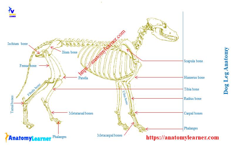
Appendicular skeleton: bones of the extremities. In this small section, we’ll briefly mention the main parts of the leg, namely the bones, muscles, and neurovasculature.Dog cephalic vein anatomy. The sacral tuber has two prominences; the cranial and caudal dorsal iliac spines. Their physical characteristics and temperaments vary from breed to breed, but they all share anatomical characteristics and exceptional, highly developed senses.Dog tongue anatomy with labeled diagram – muscle, papillae, glands, veins, and nerves, and; Dog mouth anatomy – lip, cheek, oral cavity, and salivary glands with diagram, Unique features of the dog’s mandibular body.Let’s move now forward with leg anatomy. The bones of the hindlimb are the femur, patella, fabellae (sesamoid bones), tibia, fibula, tarsus and meta-tarsus and the digital bones.
Anatomy of the Canine Knee
Anatomy of a Dog’s Paw.
- Does Mortal Kombat Gold Have A Secret Character?
- Does Warframe Support Mouse , Keyboard And Mouse Support For Ps4
- Does Jet Force Gemini Have A Split-Screen Mode?
- Donautal Geflügel Bogen : Donautal Geflügelspezialitäten (Bogen) kontaktieren
- Does Tekken 7 Have A Tier Ranking?
- Donau Apotheke Rezeptservice , E-Rezept einlösen
- Does Aer Lingus Have A Mobile Boarding Pass?
- Dog Strollers For Medium Dogs : 10 Best Dog Strollers for Hiking in 2024 — Reviews & Top Picks
- Does Samsung Gear S3 Have A 22 Mm Band?
- Dokumente Für Kindergeld – Unterlagen für die Beantragung von Elterngeld
- Does Hyrule Warriors Have A Free Patch?
- Dog Whistles On Tiktok , TikTok